IPS scientists provide cover picture for Nature Protocols, Volume 9 No. 2
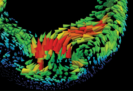
3D cell motion on a sagittal section of a mid-gastrulation Xenopus laevis embryo. Red arrows designate maximum cell speed (~4 µm min–1): mesendodermal leading-edge runners (top center); endodermal wall of inflating archenteron (left); blastopore (bottom center). Note pronounced vegetal rotation on lower right and epibolic movement throughout the periphery.

cell and tissue motion captured and visualized using flow analysis (right). (Figure: Alexey Ershov)
Reference:
J. Moosmann, A. Ershov, V. Altapova, T. Baumbach, M. S. Prasad, C. LaBonne, X. Xiao, J. Kashef, and R. Hofmann, „X-ray phase-contrast in vivo mictotomography probes new aspects of Xenopus gastrulation“ Nature (2013), doi:10.1038/nature12116
