KIT - Karlsruhe Institute of Technology image/svg+xml
Institute for Photon Science and Synchrotron Radiation
  • skip navigation
  • Home
  • Legals
  • Privacy Policy
  • Sitemap
  • KIT
    • IPS Home
    • Research
      • Research
      Research
      • Materials and Processes for Energy Technologies
      • Nanostructures, Materials & Devices for Information Technologies
      • Materials & Processes for Transport Technologies
      • Imaging Technologies for Life Science and Materials Research
      • Collaboration & Innovation
    • Methods & Facilities
      • Methods & Facilities
      Methods & Facilities
      • IMAGING Cluster
      • SCATTERING Cluster
      • SPECTROSCOPY Cluster
    • News
    • Publications
    • People
    • KIT Light Source
    • Contact
    • Jobs
    • Startpage
    • Home
    • Legals
    • Privacy Policy
    • Sitemap
    Institute for Photon Science and Synchrotron Radiation


    • Startpage
    • Methods & Facilities
    • IMAGING Cluster


    • Startseite
      • IPS Home
      • Research
      • Methods & Facilities
      • News
      • Publications
      • People
      • KIT Light Source
      • Contact
      • Jobs
    • Methods & Facilities
      • IMAGING Cluster
      • SCATTERING Cluster
      • SPECTROSCOPY Cluster
    • IMAGING Cluster
    • IMAGE
    • Methods & Instrumentation
    • X-ray Source & Optics
    • Contact

    IMAGE Beamline

    High-flux beamline for full-field hard X-ray imaging applications in materials and life sciences

    The IMAGE beamline is specifically designed for full-field hard X-ray imaging applications in materials and life sciences. The beamline prioritizes systematic in situ and operando X-ray imaging studies, along with high throughput applications (necessary for biometrics investigations in biology and paleontology) with a spatial resolution up to 1 μm. The first experimental hutch is devoted to the conduction of experiments, requiring the incorporation of specialized devices like dedicated sample environments and test chambers. The second experimental hutch houses the main experimental stations of the beamline: the tomography and the laminography stations.

    The main focus of the beamline is radiographic and tomographic imaging, both in absorption mode and in phase-contrast mode, with a resolution range from approximately 40 µm (projection radiography and tomography) down to 1 µm, covering the following fields:

    • materials research: facilities for in situ and operando characterization of electronic materials, functional materials, microsystems devices, batteries and multi-phase fluidics
    • Morphological X-ray imaging of small organisms; cell and tissue morphodynamics and organogenesis, in toto extant and fossil organisms down to organ and tissue level 

    The beamline implements various imaging modes including:

    • Contrast modes: absorption, refraction, diffraction, and fluorescence,
    • Imaging modes: radiography and microdiffraction imaging, 3D tomography and laminography

    With these modes, a variety of complementary techniques are accessible, such as:

    • White beam fast radiography and tomography in absorption contrast (distribution of the attenuation coefficients in 2D and 3D),
    • Propagation-based phase contrast imaging for low-Z and weakly absorbing materials, yielding the refractive index distribution, Element-specific imaging at absorption edges for mapping the spatial distribution of specific elements,
    • Tomography and laminography for 3D imaging of objects in absorption and phase contrast to determine the 3D distribution of the linear attenuation coefficient or the refractive index

    Methods & Instrumentation

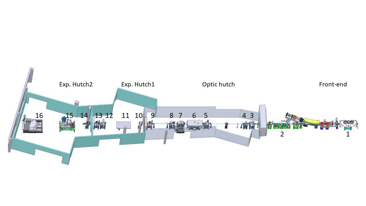
    The IMAGE beamline is located in the north-west straight-section of the storage ring. Two experimental stations are currently in operation or commisioning:


    LAMINO-II

    • In-situ – e.g., biaxial load pass experiments <10kN Large screening area  ca. 104 mm2
    • Multiple scale imaging from 200 µm down to 80 nm 3D resolution (30 nm in 2D)
    • screening of large accessible area (30 cm) and zooming into ROI with high resolution
    • Multiple contrast mechanisms (spectroscopic (XAS, XRF), phase contrasts, scattering, diffraction)

    UFO

    • UFO ('Ultra-Fast X-ray Imaging of Scientific Processes with On-line Assessment and Data-driven Process Control')
    • ultra-fast, high-resolution X-ray imaging for life and material sciences
    • non-destructive testing utilising white-beam computed tomography/laminography
    • coherent imaging by in-line holography, grating interferometry and 3D micro-diffraction imaging methods

    X-ray Source & Optics

    Characteristics

    Energy range 8 keV - 150 keV
    Energy resolution [ΔE/E] white light and optional 0.01% and 3 % bandwidth
    Source (1) Period[mm]: 51
      Number of periods: 34
      Magnetic pole gap [mm]: 18
      On-axis field amplitude [T]: 2.9
      Source size (H x V) [mm]: 1.254 x  0.065

    Layout

    Component

    Distance from the source

    High power primary slits (2)

    9 m & 10.2 m

    CVD diamond (3)

    13.18 m

    Filter unit 1 (3)

    13.387 m

    Intensity monitor 1 & Fluorescence screen 1 (3)

    14.726 m

    Filter unit 2 (4)

    14.552 m

    Secondary slits (5)

    20 m

    DMM (6)

    21.262 m

    DCM (7)

    23.737 m

    Profile monitor (8)

    24.578 m

    Intensity monitor 2 & Fluorescence screen 2 (8)

    24.863 m

    Beryllium window 1 (10)

    29.4 m

    Tertiary slits (12)

    34.6 m

    Intensity monitor 3 & Fluorescence screen 3 (12)

    34.877 m

    Beryllium window 2 (14)

    37.133 m

    sample Exp. Hutch 1 (11)

    30 m

    sample Exp. Hutch 2 (15)

    39 m

    sample Exp. Hutch 2 (16)

    44 m


    Contact(s)
    name function e-mail
    Cecilia, Angelica Deputy Head of Department, Beamline Scientist angelica cecilia ∂does-not-exist.kit edu
    Hamann, Elias Beamline Scientist, Beamline Manager elias hamann ∂does-not-exist.kit edu
    Simon, Rolf Beamline Scientist r simon ∂does-not-exist.kit edu
    last change: 2025-02-05
    KIT – The Research University in the Helmholtz Association
    • Home
    • Legals
    • Privacy Policy
    • Sitemap
    • KIT