
The application, commissioning and development of 2D and 3D X-ray imaging techniques is one of the main tasks of the Imaging group. The detector laboratory at IPS is dedicated to R&D of imaging detectors. In this lab, detectors are characterized, calibrated and tested “off-line”, independently of the synchrotron storage ring operation. Two experimental stations are available in the lab:
|
|
(1) a high-energy, high-power microfocus X-ray tube (160 kVp, 320 W) and a 5-axispositioning system mounted on an optical breadboard for moving one or more detectors and test samples independently. The X-ray system is enclosed by a fully accessible lead cabin and can be controlled remotely. The goal is to implement standardized procedures which allow qualitative and quantitative detector testing. (2) a complementary optical setup to characterize the visible light components of indirect converting X-ray detectors. The setup consists of a broad-band visible-light source, as well as a general purpose manipulator mounted on along optical rail. A photodiode is available to measure the transmission of optical components, as well as to calibrate the flux of the light source. |
Currently available detector systems
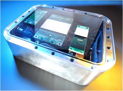
Medipix detectors:
256×256 pixels, 55 µm pixel pitch, active area 14×14 mm²
Different readout chips: Medipix2 MXR, Timepix, Medipix3
Sensor materials: Si, GaAs and CdTe
Hexa modules (matrix of 2×3 chips, active area 42×28 mm²)
Readout systems: FitPix (UTF Prague), X-KIT (IPE collaboration)
Other detectors:
Dexela 1207 (CsI scintillator, 75×75 µm² pixel size, 115×65 mm² active area)
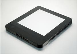
Perkin Elmer XRD 1621 flat panel (Gadox scintillator, 200×200 µm² pixel size, 400×400 mm² active area)
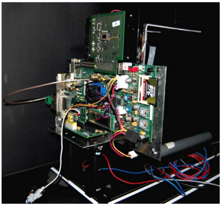
UFO camera (IPE collaboration, 2048×1088 pixels)
|
UFO camera prototype (IPE) |
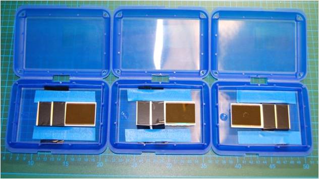
Medipix Single and Hexa assembly |
Perkin Elmer flat panel |
Applications
|
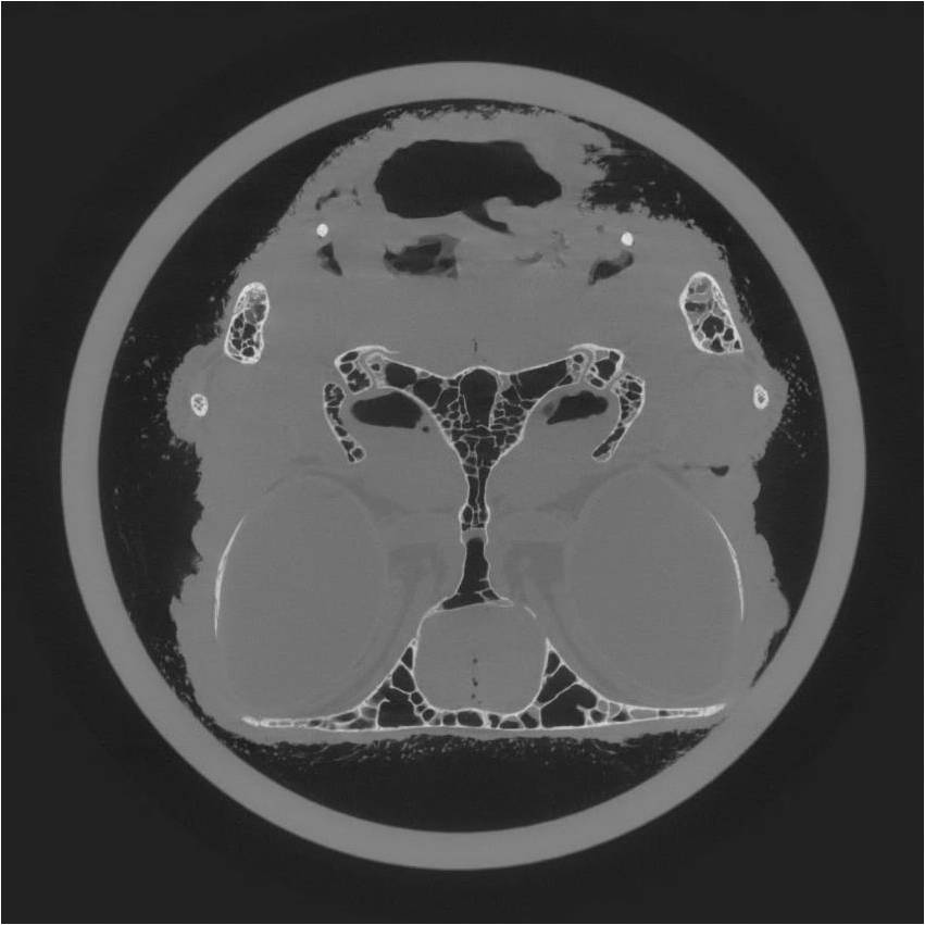
Medical / (small) animal imaging |
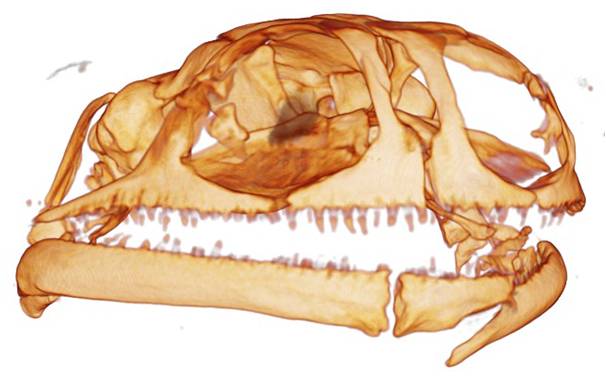
3D reconstruction / rendering |
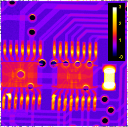
Material testing / quality assurance |
|
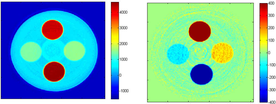
„Spectroscopic“ CT |
||
Current lab upgrade
Upgrade plans:
- New mechanics for the X-ray tube
- Add-on for Grating Interferometry (method transfer synchrotron ↔ lab)
- Integration of DESY‘s high speed Medipix3 readout „LAMBDA“
- Spectroscopic CT in combination with Grating Interferometry
- Upgrade of the visible light setup
|
Recently delivered GaAs and CdTe Medipix3 Hexa assemblies for LAMBDA readout |
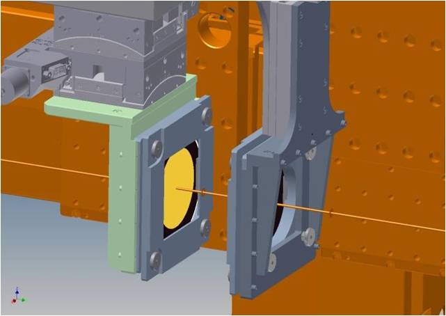
Grating Interferometry add-on |
Collaborations
IPS is part of several collaborations like SCINTAX, Galapad,UFO, the HGF detector initiative, and the Medipix3 collaboration, which all aim for developing and improving X-ray imaging detector systems.
BIRD: Scintillator and semiconductor based imaging detectors
- FMF Freiburg
GALAPAD: Development of GaAs sensors and pixel detectors
- DESY
- TSU Tomsk
- FMF Freiburg
HiZPad / HiZPad2: High Z pixel detectors
- Synchrotrons: ESRF, DESY, ELETTRA, DIAMOND, SLS, SOLEIL
- CPPM, RAL
- FMF Freiburg, University of Surrey
- Dectris
EDAS: CdTe pixel detectors
- FMF Freiburg