Dose-efficient X-ray method allows imaging of dynamic processes in living organisms over extended time periods
- Date: Dec 2023
Dose-efficient X-ray method allows imaging of dynamic processes in living organisms over extended time periods
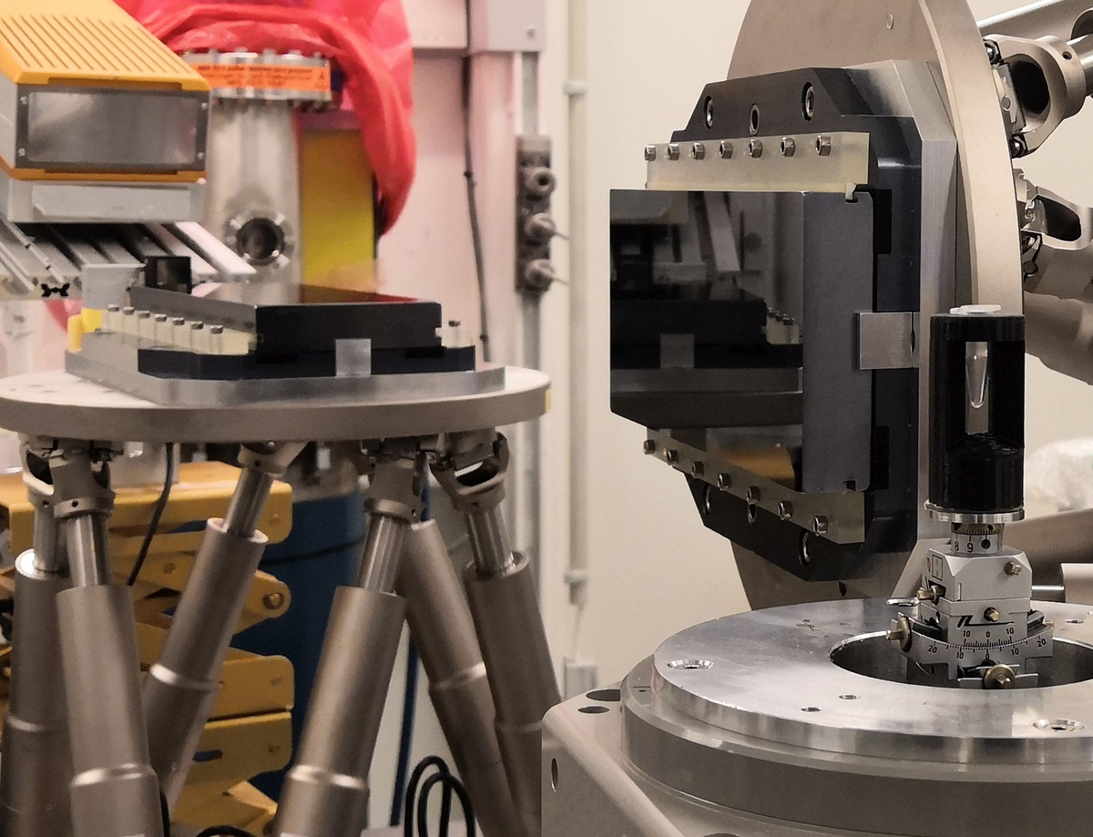
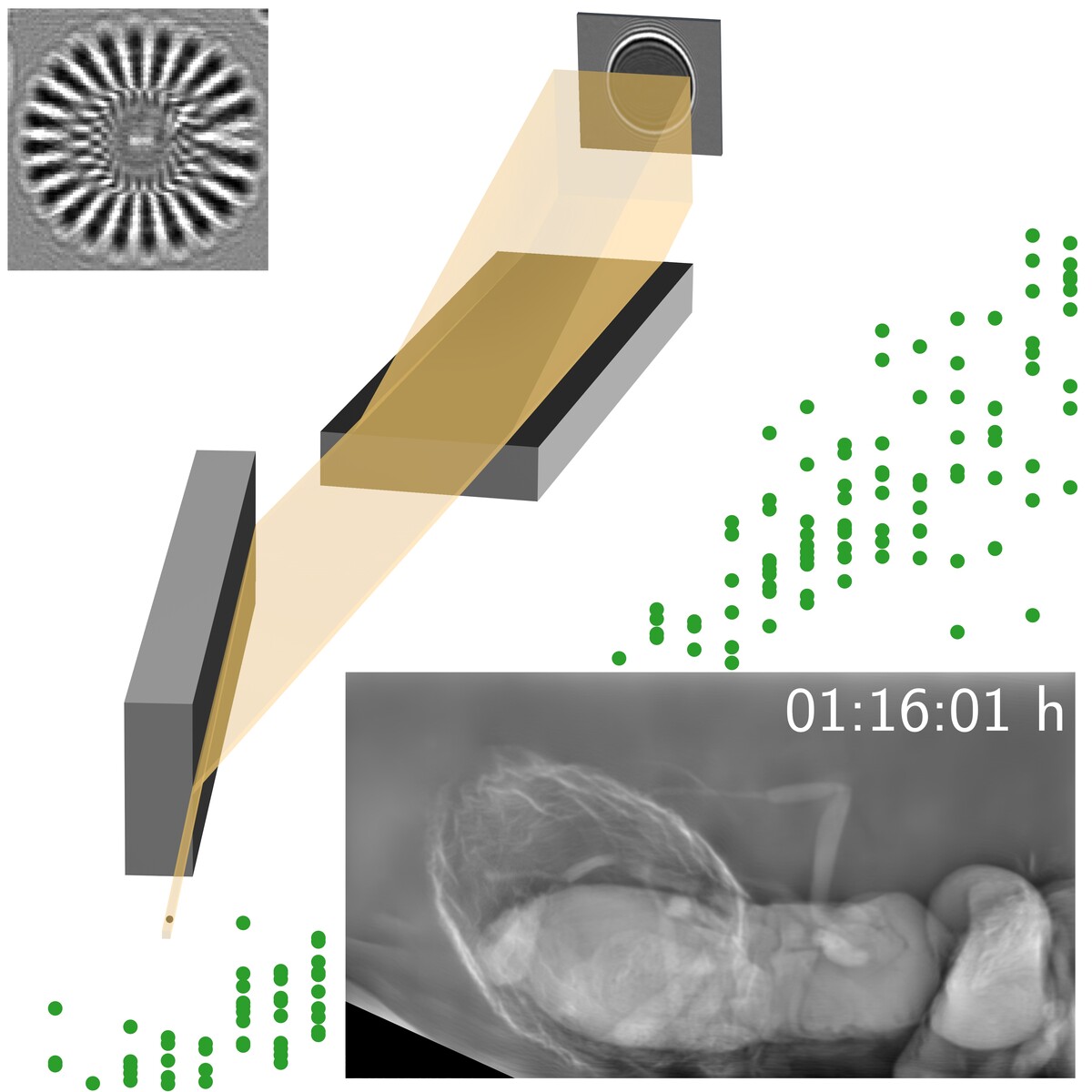
X-ray imaging of living samples is challenging due to the high radiation load deposited in the specimens, which is required to obtain sufficient image quality, especially at high resolution. At IPS, a method has been developed to achieve high dose efficiency by combining X-ray phase contrast with a Bragg magnifier and a highly-efficient single-photon-counting detector.
The developed Bragg magnifier consists of two silicon crystals that magnify the X-ray wavefield behind the sample by asymmetric Bragg diffraction. In this way, the high detection efficiency of single-photon-counting detectors can be exploited while maintaining micrometer resolution. The method allows for extended observation times for in vivo or in situ studies of dose-sensitive samples before the onset of radiation damage.
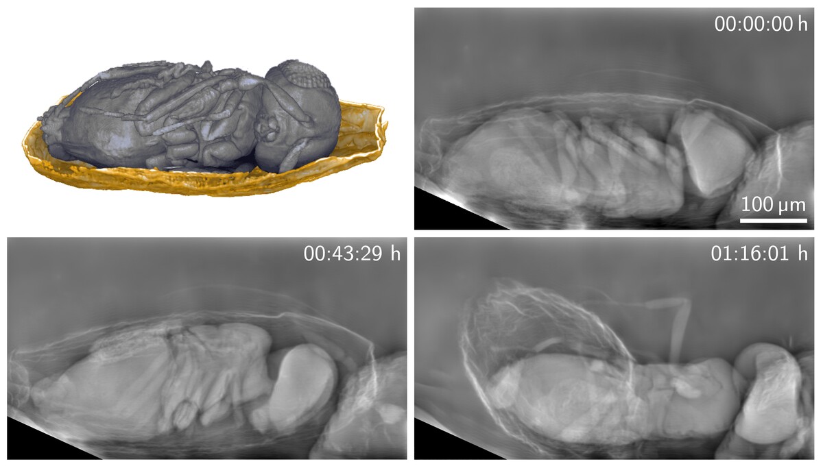
As a pilot application, living parasitoid wasps were observed for more than 30 minutes inside their host eggs before and during emergence, revealing the flexible biting capability of their mandibles while perforating the eggshell and the involvement of different body parts during emergence.
The method has been developed in the frame of the BMBF projects CODE-VITA and HIGH-LIFE.
Further information
- KIT press release (in German)
- Optica news release
- Optics and Photonics News
- Interview with AZO Optics
Original publication: R. Spiecker, P. Pfeiffer, A. Biswal, M. Shcherbinin, M. Spiecker, H. Hessdorfer, M. Hurst, Y. Zharov, V. Bellucci, T. Farago, M. Zuber, A. Herz, A. Cecilia, M. Czyzycki, C. S. Baraldi Dias, D. Novikov, L. Krogmann, E. Hamann, T. van de Kamp, T. Baumbach, “Dose-efficient in vivo X-ray phase contrast imaging at micrometer resolution by Bragg magnifiers,” Optica 10, 1633-1640 (2023). https://doi.org/10.1364/OPTICA.500978