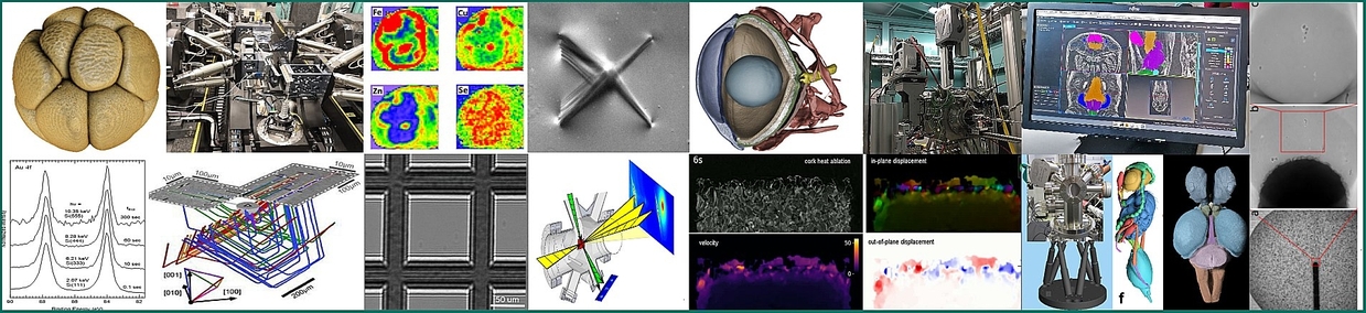
IMAGING Cluster: Instrumentation & Methods for Multiscale X-ray Imaging in Materials Science & Life Science
The IMAGING Cluster includes advanced imaging beamlines and related laboratory infrastructure at KIT Synchrotron, as well as experimental stations at other prominent synchrotron facilities like the European Synchrotron Radiation Facility (ESRF, Grenoble) and PETRA III in Hamburg. The available infrastructure and methodical portfolio play a central role for the cluster's mission to carry out innovative research in advanced imaging across various scientific fields and applications.
| Beamlines, Experimental Stations and Labs |
|
|
- at KIT: Lamino II, UFO I & II, Cryo-MIC, WBTop
- at other facilities: Lamino I@ESRF, HIKA@PETRA III
- 3 Mobile set-ups: XDL, BMM, BMC
- Offline Lab. Infrastructures: BIO Lab., CT/CL Lab., Detector Test Lab.
|
| Laboratory laminography / tomography |
UFO technology for fast and HT imaging |
- Transfer between SR and lab technology
- high variability in contrast µ, GI, spectroscopic imaging
|
- UFO I and UFO II stations - up to 20 CT-scans/sec,
- High throughput (up to 1500 specimen / 2 days)
- On-line control and reconstruction
|
| Synchrotron laminography / tomography |
Microscopy |
- Lamino I @ ESRF for high coherence applications
- Lamino II Operando, multiple contrast
|
- BioMiC: FZP - UHV-Cryo; 5-12 keV 30 nm 2D
- MIQA @ PETRA III 80 nm 3D SOC, 6nm step-scans
|
| The IMAGING Cluster allows morphological full-field 2D, 3D and 4D imaging in materials research and life sciences based on: |
X-ray optical techniques, beam conditioning options, for example: |
- instrumentation and methods for multiple-scale imaging from >200 µm down to <100 nm (3D) (30 nm in 2D)
- contrast mechanisms: phase (differential F via GI, DEI, Zernike, PBI/ holographic imaging, BMI), spectroscopic (XANES, XRF), dark-field, Bragg / Laue diffraction
- screening of large accessible area and zooming into ROI with high resolution
- high throughput > 750 specimen/day; ultrafast imaging at 20 CT/s
- white, pink, monochromatic beam conditions
|
- 2D Parallel & conic beam projection modes
- 2D X-ray microscopy modes (TXM, SXM)
- 3D tomography, laminography, conv., limited angle, spiral
- Dynamic imaging: 3D cine-radiography, 4D cine-tomography
|