KIT - Karlsruhe Institute of Technology image/svg+xml
Institute for Photon Science and Synchrotron Radiation
  • skip navigation
  • Home
  • Legals
  • Privacy Policy
  • Sitemap
  • KIT
    • IPS Home
    • Research
      • Research
      Research
      • Materials and Processes for Energy Technologies
      • Nanostructures, Materials & Devices for Information Technologies
      • Materials & Processes for Transport Technologies
      • Imaging Technologies for Life Science and Materials Research
      • Collaboration & Innovation
    • Methods & Facilities
      • Methods & Facilities
      Methods & Facilities
      • IMAGING Cluster
      • SCATTERING Cluster
      • SPECTROSCOPY Cluster
    • News
    • Publications
    • People
    • KIT Light Source
    • Contact
    • Jobs
    • Startpage
    • Home
    • Legals
    • Privacy Policy
    • Sitemap
    Institute for Photon Science and Synchrotron Radiation


    • Startpage
    • Methods & Facilities
    • IMAGING Cluster


    • Startseite
      • IPS Home
      • Research
      • Methods & Facilities
      • News
      • Publications
      • People
      • KIT Light Source
      • Contact
      • Jobs
    • Methods & Facilities
      • IMAGING Cluster
      • SCATTERING Cluster
      • SPECTROSCOPY Cluster
    • IMAGING Cluster
    • HIKA
    • Layout & Characteristics
    • Contact

    HIKA: The Hierarchical Imaging KArlsruhe Station at PETRA III

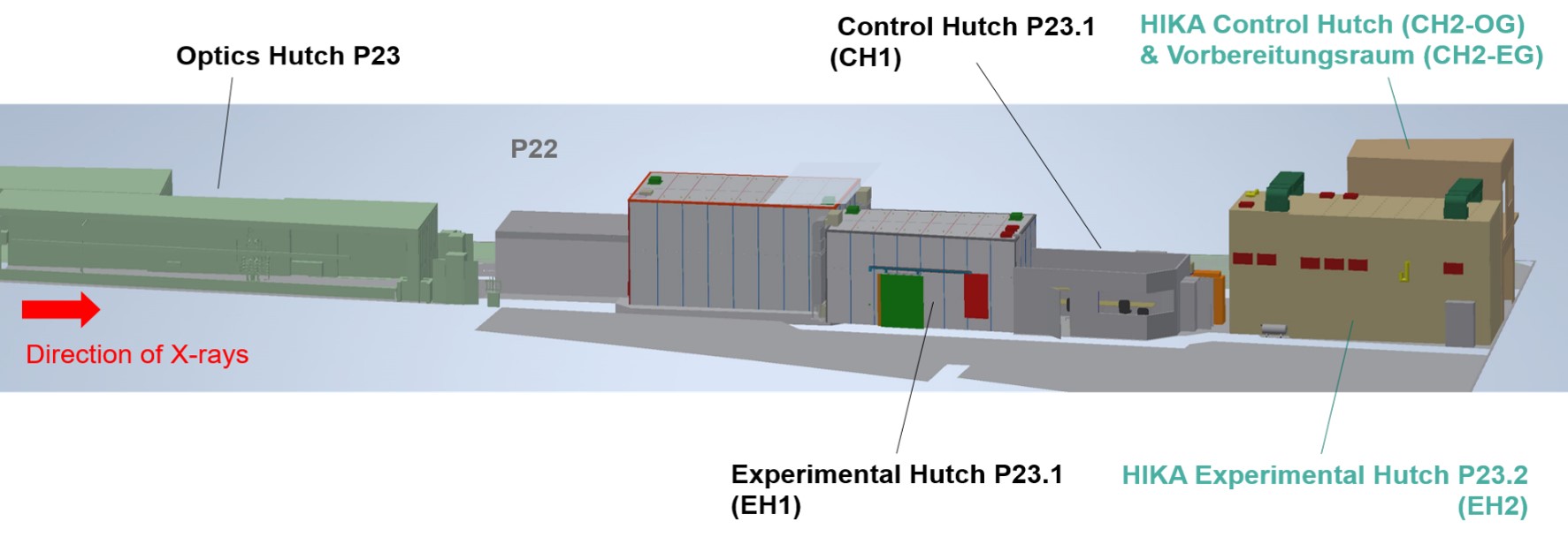
    The Hierarchical Imaging KArlsruhe Station is currently under construction at the In situ X-ray Diffraction and Imaging Beamline P23 at PETRA III. HIKA will offer a portfolio of morphological full-field imaging methods, including:

    • micro-tomography & laminography for 3D morphology 
    • high-throughput large comparative studies
    • operando, in situ and in vivo imaging for 4D morphodynamics
    • combined BMI, parallel beam imaging, and X-ray microcopy 
    • multiscale and hierarchical X-ray imaging  
    • multiple X-ray contrasts, light microscopy for correlated imaging

    HIKA will offer various operational modes, with either a combination of, or fast switching between:
     

    • parallel beam imaging: absorption, phase contrasts (grating interferometry, propagation based up to 4 m, Bragg Magnifiers for large FOV)
    • microscopy: full-field TXM & SXRM (can be equipped with up to 4 independent optical elements, switchable resolution, sample scanning precision < 6 nm)
    • 3D imaging modes: high resolution CL & CT, nominally < 80 nm sphere of confusion
    • dose-efficient imaging: Single distance + up to 31 keV Bragg Magnifier Optics + SPC-Detectors


    and will implement serial CT with smart, standardized morphological DAQ and DA protocols suitable for large, comparative studies, including the development and application of:
     

    • smart robotics and algorithms for large sample series and for control of FOV, ROI, resolution, CNR and SNR to ensure image quality and data comparability between different specimens, sample series and imaging modes 
    • smart communication between the DAQ instrumentation and AI-based image analysis pipelines
    • fully automated pipelines for digitizing morphology and morphodynamics and correlation 
    • AI-based, automated 3D & 4D morphological imaging for systematic, large scale applications 


    Beamline Layout

    The P23.HIKA extension is located on the P23 beamline in the PETRA III experimental hall “Ada Yonath”

    HIKA Contact(s)
    name function e-mail
    Sato Baraldi Dias, Carlos Beamline scientist in charge of the HIKA station carlos dias ∂does-not-exist.kit edu
    Czyzycki, Mateusz Beamline Scientist mateusz czyzycki ∂does-not-exist.kit edu
    last change: 2024-12-12
    KIT – The Research University in the Helmholtz Association
    • Home
    • Legals
    • Privacy Policy
    • Sitemap
    • KIT