KIT - Karlsruhe Institute of Technology image/svg+xml
Institute for Photon Science and Synchrotron Radiation
  • skip navigation
  • Home
  • Legals
  • Privacy Policy
  • Sitemap
  • KIT
    • IPS Home
    • Research
      • Research
      Research
      • Materials and Processes for Energy Technologies
      • Nanostructures, Materials & Devices for Information Technologies
      • Materials & Processes for Transport Technologies
      • Imaging Technologies for Life Science and Materials Research
      • Collaboration & Innovation
    • Methods & Facilities
      • Methods & Facilities
      Methods & Facilities
      • IMAGING Cluster
      • SCATTERING Cluster
      • SPECTROSCOPY Cluster
    • News
    • Publications
    • People
    • KIT Light Source
    • Contact
    • Jobs
    • Startpage
    • Home
    • Legals
    • Privacy Policy
    • Sitemap
    Institute for Photon Science and Synchrotron Radiation


    • Startpage
    • Methods & Facilities
    • SPECTROSCOPY Cluster


    • Startseite
      • IPS Home
      • Research
      • Methods & Facilities
      • News
      • Publications
      • People
      • KIT Light Source
      • Contact
      • Jobs
    • Methods & Facilities
      • IMAGING Cluster
      • SCATTERING Cluster
      • SPECTROSCOPY Cluster
    • SPECTROSCOPY Cluster
    • SUL-X
    • Methods
    • Layout
    • Characteristics
    • Contact

    SUL-X Beamline


    The X-ray beamline of the Sychrotron Laboratory for Environmental Studies (Synchrotron Umwelt-Labor) combines diffraction, absorption and fluorescence measurements on environmentally-relevant materials with microfocusing capabilities.
     

    Sources of pollution, path of contaminants, and the application of synchrotron radiation-based techniques via Molecular Environmental Science (MES) to environmental questions are the main tasks of this beamline.
     

    The SUL X beamline is major part of the Synchrotron Laboratory for Environmental Studies supported by the German governments (BMBF and state Baden Württemberg) for dealing environmental questions with synchrotron radiation methods. Therefore this beamline is designed for combined measurements with a microfocusing capability.
     

    Environmental samples are generally complex (e.g., contaminated soils, mining dump sediments), consisting of mixtures of mineral phases (amorphous, crystalline, with micrometer or nano-scale particle sizes), microbes, and in some cases vegetable material. Spatial distribution, speciation, and phase association of trace levels of contaminants are the basis for risk assessment and development of remediation strategies (e.g. arsenic speciation and distribution in groundwater related sediments). This problem is best addressed by synchrotron based techniques capable of providing spatial resolution in the micrometer range while allowing the combination of microfocused techniques (XRF, XAS, XRD, IR), as foreseen in the SUL concept.

    Methods

    The SUL X-ray beamline closes the spectral gap between soft and hard X-ray spectroscopy and allows investigation of samples - without remounting them - sequentially with the following methods and modes:

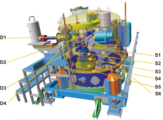
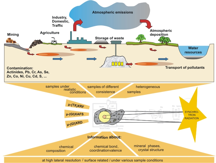
    Fluorescence Spectroscopy:

    •     elemental mapping of an extended sample :e.g. thin section of a contaminated soil sample
          (microfocused beam size mode)
    •     high sensitivity to low concentrations (primary “white light” beam with its high flux mode)

    Absorption spectroscopy for all elements between S(Al) and U:

    •     information about the local atomic geometry (EXAFS)
    •     chemical state of the absorbing atom (XANES)
    •     investigations on ordered (crystalline) and disordered (amorphous, liquid) materials
    •     dilute species and light elements (fluorescence mode of XAS)

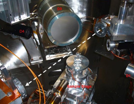
    Diffraction experiments (powder and aggregates of crystals):

    •     location of pollutant atoms within a crystalline mineral matrix
    •     investigation of sample concentrations, chemical states of elements and their
          associated mineral phases down to the μm scale
    •     essential key parameters for environmental and health risk assessment

    Advantages

    •     Nondestructive, Surface / volume sensitive
    •     Three X-ray techniques with microfocus without sample remounting
    •     Spectroscopy from light elements S(Al) to U at a single beamline

    Purpose, summarized

    •     Environmental science, material science, biology
    •     Focus on inhomogeneous, complex samples
    •     Spatial element distribution, speciation, and mineral phase determination

    Layout

    Overview

    Front-End with first, horizontal, focusing mirror (M1) First part of the optic hutch with double crystal monochromator (DCM) and second, vertical, focusing mirror (M2) End of the optic in experimental hutch with the Kirckpatrick -Baez mirror system (KBM)

    Experimental station - 3D model


     Detectors

      Detector Type Main specifications

     D1

     Fluorescence

    7 element Si(Li) (Gresham, now e2v)

    Resolution (averaged over all elements, optimum peaking time):
    <140 eV (at 5.9 keV, 1000 cps)
    <310 eV (at 5.9 keV, 100.000 cps)

    Area per element 30 mm2

     D2

     Absorption

    3 ionisation chamber (Oxford Instruments, IC-Plus type)

     Active lenght 5cm, Kapton windows 6 µm thick

     D3

     Diffraction

    CCD detector (Photonic science)

    Active area 80x120mm2 (hor. x vert.)

    Fiber optic 3.46:1 scaling down pixels 2048x2048, 16 bit dynamic through fusion mode

    Readout time 3.3s – 21s
    Vacuum compatible

     D4

     Selection of sample spots

    Optical microscope (TSO Spezialoptik)

    Resolution 2µm,

    Transmitted and reflected light, Vacuum compatible

    Sample stage

     Movement

     Range

     S1

     z stage

     105 mm

     S2

     x/y stage

     100mm / 40mm

     S3

     phi circle

     360°

     S4

     chi cradle

     10°

     S5

     theta circle

    360°

     S6

     2 theta circle

      -160° to +15°


    7 Element Si(Li) fluorescence detector (Gresham)

     
    In-vacuum CCD detector (Photonic Science)

    Key parameters of the beamline


    Energy range

    2.3 kev - 19 keV (S K-edge, U L3-edge), later 1.4 keV (Al K-edge)

    Energy resolution (DE/E)

    Si(111) 2x10⁻⁴; Si(311) 1x10⁻⁴; YB66(004) 5x10⁻⁴;


    (theoretical values) 

    Source

    Wiggler (27 pole each 74 mm), later upgrade undulator (100 pole each 14mm)

    Optics (sorted in beam direction)

    Toroidal mirror, horizontally focusing, vertically collimating

    DCM with Si(111), Si(311), YB66(400) and mirror (white light)

    Cylindrical mirror with three coatings, low energy band path, vertically focusing

    Precise slit in focus, defining a “new” source with adjustable size

    Elliptical Kirkpatrick Baez mirror system, focusing “new” source

    Monochromatic or “white light” beam path, selectable.

    Beam size at sample position

    1 mm (hor) x 1mm (vert) down to 30 µm x 25 µm

    (later down to 10 µm x 10 µm)

    Flux at sample position

    4 x 10¹⁰ (5keV), 3 x 10¹⁰ (10kev), 3 x 10¹⁰ (20keV) ph/s / 100mA e- current in spot size (FWHM) 0.2 mm (hor) x 0.1 mm2

    Experimental station


    Sample diffractometer with theta, phi circle and chi cradle (10°), xyz linear stages

    CCD detector on 2theta arm for diffraction

    3 retractable ionization chambers for absorption

    7 element Si(Li) fluorescence detector for fluorescence

    Optical microscope

    Diffractometer and detectors all in vacuum vessel

    Sample environment

    Chambers for different environments (gases, liquids, T-controlled) are planned; low temperature environment down to -5 °C (small samples, peltier cooled), and down to -20 °C (flat samples, N 2 cryostream) available.


    Contact(s)
    name function e-mail
    Göttlicher, Jörg Beamline Scientist joerg goettlicher ∂does-not-exist.kit edu
    Steininger, Ralph Deputy Head of Department, Beamline Scientist ralph steininger ∂does-not-exist.kit edu
    last change: 2022-12-07
    KIT – The Research University in the Helmholtz Association
    • Home
    • Legals
    • Privacy Policy
    • Sitemap
    • KIT