
NOVA

NOVA – Network for Online Visualization and Synergistic Analysis of Tomographic Data
Synchrotron X-ray microtomography offers unique opportunities for the morphological analysis of animals. Internal structures become observable even in opaque organisms in a non-invasive, three-dimensional way, and with sub-micron resolution. The method thus allows for the investigation of objects from a wide field of biological research, from biomechanics, dealing with the complex dynamics of muscle-skeleton interactions, neurobiology with the topology of sensory centres in the brain, to developmental biology, with differential comparisons of organism growth stages. So far the enormous potential of collaborative work on the same specimens is mostly untapped. By using a new collaborative approach, NOVA aims to create new possibilities that allow for a more efficient use of the valuable beam time at tomographic synchrotron beamlines through the coordination of research on different organ systems and a regulation of the data usage by a common data policy.
The (associated) biological partners of the joint research project NOVA cover different scientific aspects of morphology and thus serve as a model community for the cooperative, integrative approach. As exemplification for the biological objects, complementary aspects of insect head morphology will be investigated. On this basis, a tomography catalogue (and most notably a freely accessible one) will be created in cooperation with the operators of the tomography instrumentation at PETRA III. Additionally, datasets already obtained at ANKA in the framework of ASTOR will be added. In a later stage of the project additional scientific communities plan to be enlisted to participate in this open and cooperative way of data usage.
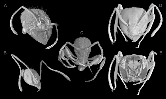
Examples of ant species recorded during the first NOVA beamtime in October 2016. A, B - Pheidole lucretii. Comparison of soldier (A) and worker morphotype (B) of the same species. C - Gnamptogenys striatula. D, E - Lasius fuliginosus. (D) is an anterior overview, whereas (E) is a virtual cross section of the ant head showing the central nervous system (CNS; black arrow head), and muscles of the mandibles (white arrow head). From: Schmelzle, S., Heethoff, M., Heuveline, V., Lösel, P., Becker, J., Beckmann, F., Hammel., J.U., Kopmann, A., Mexner, W., Vogelgesang, M., Tan Jerome, N., Betz, O., Beutel, R., Wipfler, B., Blanke, A., Harzsch, S., Baumbach, T., van de Kamp, T., “The NOVA project - maximizing beam time efficiency through synergistic analyses of SRμCT data”, Proceedings of SPIE (2017), in press.