
ARTHROPODS
Arthropod imaging @ IPS & LAS
Constituting an estimated 80% of all living animals, arthropods are by far the most species-rich phylum of organisms. They include insects, spiders, crustaceans and a number of smaller groups, all characterized by an exoskeleton, a segmented body and paired appendages. Despite their evolutionary success and vast diversity, many aspects of arthropod biology are still unknown.
X-ray imaging is highly suited to visualizing internal morphological characteristics of opaque millimeter-sized specimens, especially those which are not dissectible or are of considerable value: in particular, synchrotron-based X-ray imaging is capable of providing real-time three-dimensional information on arthropod morphology, thus stimulating advances in a wide range of research fields ranging from evolutionary biology and paleontology to functional morphology, morphodynamics and biomimetics.
An interdisciplinary network of international partners from universities, research centers and museums, scientists from KIT’s Laboratory for Applications of Synchrotron Radiation (LAS) and Institute for Photon Science and Synchrotron Radiation (IPS) investigate arthropod structure and function using a variety of different X-ray imaging techniques such as (fast) tomography, laminography, radiographic movies and in vivo cine-tomography. Apart from the scientific questions directly relevant to the organisms themselves, arthropods also serve as important test samples for the development and evaluation of biological X-ray imaging techniques both for conventional X-ray sources and synchrotron radiation facilities.
Functional morphology & Biomimetics
Arthropod joints are difficult to examine by traditional methods such as light or scanning electron microscopy, but modern X-ray imaging now offers the possibility to analyze their structure and function in three dimensions. When, in 2011, we described the first biological screw-and-nut type joint in the hips of weevils [1], a beetle family with over 60,000 recorded species, it became apparent how little is still known about insect functional morphology. With millions of species, insects constitute a rich, but still largely unexploited source of biological joint systems, of which some may be suitable role models for biomimetic design.
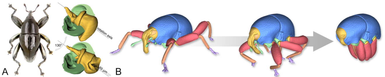
Examples taken from recent studies on beetle functional morphology. A. Screw-and-nut type hip joint in a weevil [1]. B. Interactive animated 3D reconstruction of a weevil’s defensive movements, which include unique mechanical blockings of head and legs [2].
In a recent study in collaboration with the Bio-inspired Photonics Group of the University of Cambridge (to investigate structural colors in dock leaf beetles (Gastrophysa viridula). We employed traditional morphological techniques (such as TEM), fast synchrotron-based X-ray microtomography and optical microspectroscopy in order to identify morphology, composition and optical appearance of the bio-photonic structures and to study the development of the cuticular reflector [3].
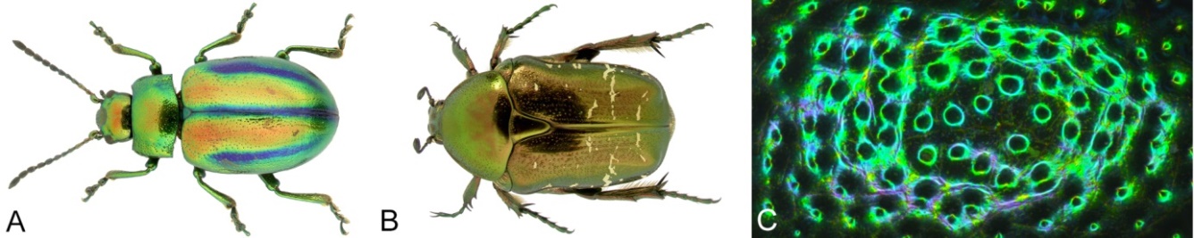
Examples for structural colors in beetles. A. Chrysolina fastuosa. B. Cetonia aurata. C. Detail of the elytron of Gastrophysa viridula.
In a recent collaboration with architects from the University of Stuttgart, a biomimetic research pavilion based on beetle elytra was constructed in order to evaluate fiber composites for building structures [4]. This project demonstrated how biological role models can influence architectural design and received a lot of media attention, including a TV documentary for the ARTE channel (“Insekten, Superhelden auf sechs Beinen”).
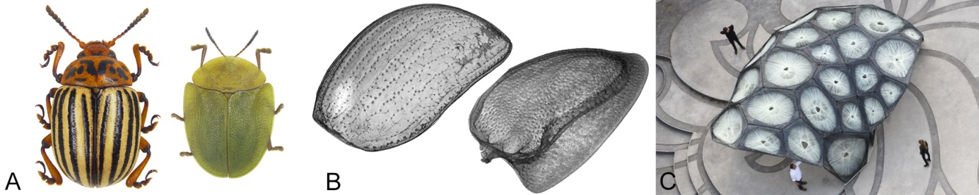
A biomimetic research pavilion based on beetle elytra [10]. A. Potato beetle and Green tortoise beetle. B. Transparent volume renderings of isolated elytra of both species. C. Final pavilion at the University of Stuttgart.
When studying 30-million-year old mineralized histerid beetles from the fissure fillings of the Quercy region in France, we found that insects from non-amber collections may also contain detailed internal anatomical characters, thus allowing species description and phylogenetic analysis as done for extant specimens [3]. Our findings suggest that mineralized insects constitute a comprehensive but as yet largely neglected source for three-dimensional anatomical data, therefore providing a huge potential for important new discoveries.
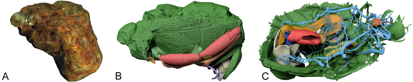
Digital reconstruction of a mineralized histerid beetle from Paleogene fissure fillings [3]. A. Photograph of the fossil. B. Beetle digitally isolated from stone matrix. C. Perspective view of the preserved internal anatomy including tracheal network, alimentary canal and genitals.
References
[1] van de Kamp, T., dos Santos Rolo, T., Baumbach, T. & Krogmann, L. (2014): Scanning the past – synchrotron X-ray microtomography of fossil wasps in amber. Entomologie heute 26: 151-160.
[2] Riedel, A., dos Santos Rolo, T., Cecilia, A. & van de Kamp, T. (2012): Sayrevilleinae Legalov, a new subfamily of fossil weevils (Coleoptera, Curculionoidea, Attelabidae) and the use of synchrotron microtomography to examine inclusions in amber. Zoological Journal of the Linnean Society 165: 773-794.
[3] Schwermann, A.H., dos Santos Rolo, T., Caterino, M.S., Bechly, G., Schmied, H., Baumbach, T. & van de Kamp, T. (2016): Preservation of three-dimensional anatomy in phosphatized fossil arthropods enriches evolutionary inference. eLife 5: e12129.