
CODE-VITA
CODE-VITA is a joint project between the University of Heidelberg and KIT (Laboratory for Applications of Synchrotron Radiation) to develop synchrotron measurement technology for dose-efficient X-ray imaging, and to apply this exemplarily for research into development processes in model organisms which are optically opaque and therefore less accessible to visible light optical methods.
Central topics of the project are metrological developments to realise:
- beam expansion, since the extremely small emittance, small source size and divergence of synchrotrons of the third and fourth generation at the sample location result in excellent brilliance and coherence properties, but on the other hand, with conventional beamline lengths, they result in limited beam cross sections and a limitation of the field of view (FoV). For this purpose, substantial two-dimensional beam expansion should be achieved while largely retaining beam coherence.
- substantially increased dose efficiency, which is a major problem in particular for biological imaging at synchrotron sources of the highest brilliance. For this purpose, the aim is to create experimental conditions for high contrast-to-noise ratios (CNR) and high detection efficiency by means of suitable instrumental developments and the integration of suitable reconstruction algorithms.
There are two experimental approaches:
|
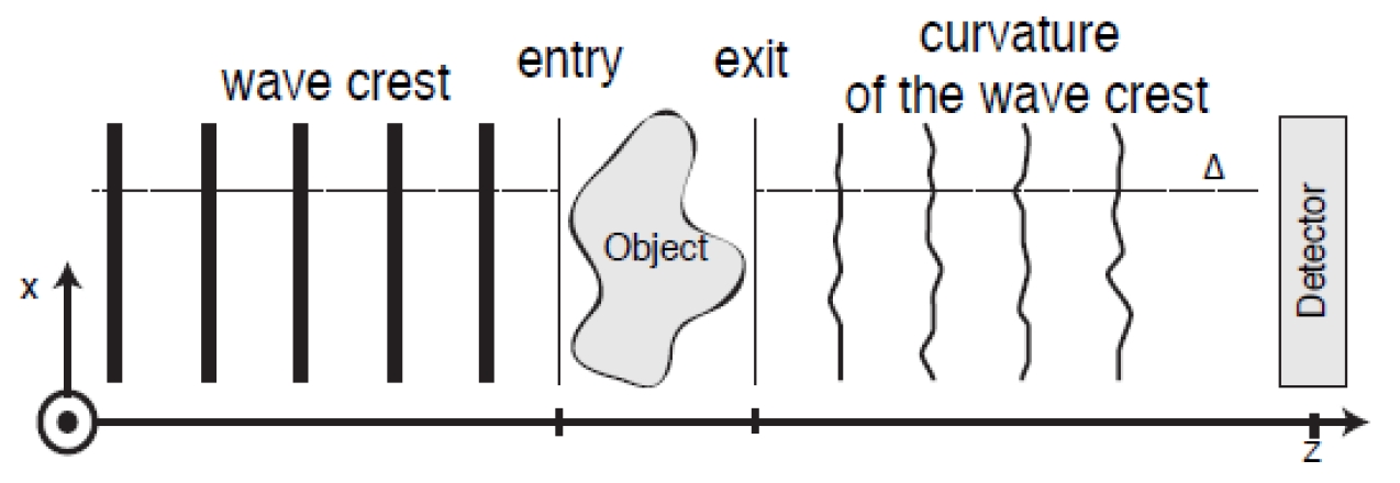
1) Propagation-based phase-contrast imaging based on novel single-distance quasi-particle phase retrieval algorithm for large z and strong phase objects [1]. Larger propagation distances increasingly release information at high frequencies at the same dose. |
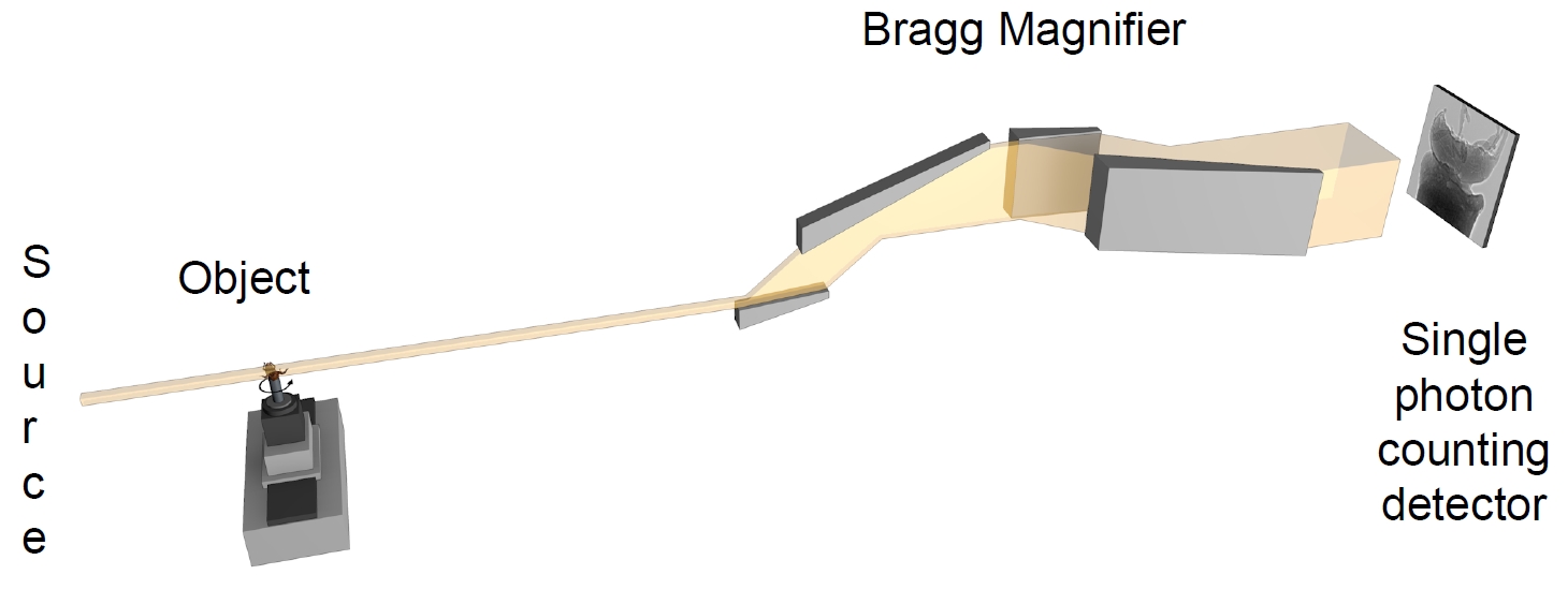
2) Bragg Magnifier crystal optics utilise asymmetric Bragg diffraction in single crystals (Si, Ge) to magnify the X-ray beam up to x200 at ~30 keV while retaining high coherence. |
|
In combination with single photon counting detectors, this will allow dose-efficient imaging at pixel sizes down to 0.3 μm for in vivo imaging during developmental processes. Alternatively a second option using a Bragg Magnifier Conditioner (BMC) to expand the X-ray beam up to 6×6 cm² allows for imaging of entire organisms with high coherence (so-called beamline extender). |
|
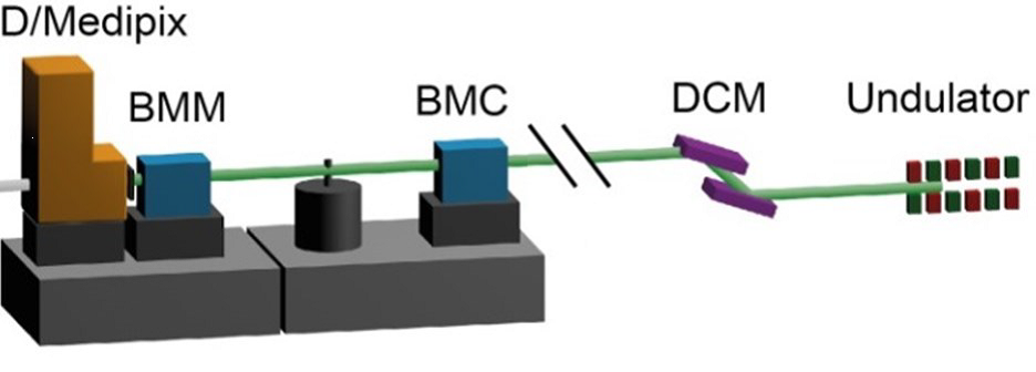
To realize these experiments the HiKA experimental station at PETRA III will be a key element. The setup will comprise Bragg Magnifier modules and a propagation-based phase contrast imaging module based on conventional scintillators and CCD/CMOS cameras.
CODE-VITA is funded by the BMBF under grant numbers 05K16VH1 (U. Heidelberg) and 05K16VK6 (KIT)
Publications
- Capillarity and active cell movement at mesendoderm translocation in the Xenopus gastrula”, Nagel, M.; Barua, D.; Damm, E. W.; Kashef, J.; Hofmann, R.; Ershov, A.; Cecilia, A.; Moosmann, J.; Baumbach, T.; Winklbauer, R., 2021. Development <Cambridge>, 148 (18)
- “Introducing Biomedisa as an open-source online platform for biomedical image segmentation”, Lösel, P., van de Kamp, T., Jayme, A. et al., Nat Commun 11, 5577 (2020).
- “Quantitative morphometric analysis of adult teleost fish by X-ray computed tomography”, Weinhardt, V., Shkarin, R., Wernet, T. et al., Sci Rep 8, 16531 (2018).
- “3D biodegradable scaffolds of polycaprolactone with silicate-containing hydroxyapatite micro-particles for bone tissue engineering: high-resolution tomography and in vitro study”, Shkarina, S., Shkarin, R., Weinhardt, V. et al., Sci Rep 8, 8907 (2018).
- “Phase retrieval for arbitrary Fresnel-like linear shift-invariant imaging systems suitable for tomography”, S. Hrivňak, A. Hovan, J. Uličný, and P. Vagovič, Biomed. Opt. Express 9, 4390-4400 (2018).
- “Fast Fresnel propagation through a set of inclined reflecting planes applicable for X-ray imaging”, S. Hrivňak, J. Uličný, and P. Vagovič, Opt. Express 26, 34569-34579 (2018).
- “Fresnel diffractograms from pure-phase wave fields under perfect spatio-temporal coherence: Non-linear/non-local aspects and far-field behavior”, Trost, F., Hahn, S., Müller, Y. et al. Sci Rep 7, 17706 (2017)