
X-REGIO
X-REGIO forms an interdisciplinary consortium of Russian and German research institutions bringing together developers of X-ray techniques (X-ray imaging, computation, and imaging analysis) and research groups with strong interest in systematic use of the methodical ensemble, in particular for 3D multi scale imaging of tissues and cells in scaffolds and model organisms for the application fields of
- Regenerative biology
- Functional Genomics
- Nanotoxicology
All these applications face similar challenges so that they all demand for the development of hierarchical imaging.
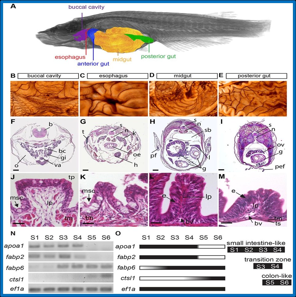
The medaka intestinal tract shows morphological and functional homology to the mammals. (A) 3D image of an adult medaka taken by x-ray microCT. Anatomical landmarks of the gut are highlighted. Data were used for (B) reconstruction of the lumen of the buccal cavity. (C) esophagus, B, C rostral to caudal perspective). (D) midgut; anterior: left with densely packed folds; posterior: right with elongated folds. (E) posterior gut; anterior: left; posterior: right. (F-I) H&E stained transverse sections of adult gut along rostro-caudal axis. Histology of intestinal folds in each segment in (FI) is shown in (J-M). Morphology of the folds varies along the rostro-caudal axis. Identification, visualization and clonal analysis of intestinal stem cells in fish. From: Aghaallaei, N.; Gruhl, F.; Schaefer, C. Q.; Wernet, T.; Weinhardt, V.; Centanin, L.; Loosli, F.; Baumbach, T.; Wittbrodt, J., 2016. Development <Cambridge>, 143 (19), 3470–3480. doi:10.1242/dev.134098
X-REGIO is geared towards multi-scale X-ray imaging from small complete animal scanning, e.g. of Zebra fish series 10μm resolution by phase contrast techniques (such as grating interferometry and analyzer-based imaging), single-distance holographic phase tomography and laminography, as well as sub-micron scanning of region of interests (ROIs). New hard full-field X-ray microscopic methods will push the limits of spatial resolution available at ANKA down to a few ten nanometres.
Publications
- Aghaallaei N., Gruhl F., Wernet T., Weinhardt V., Loosli F., Baumbach T., and Wittbrodt J., Identification, visualization and clonal analysis of intestinal stem cells in fish, The Company of Biologists: Development, revised version submitted (2016).
- Takamiya, M., Xu, F., Suhonen, H., Gourain, V., Yang, L., Ho, N.Y., Helfen, L., Schröck, A., Etard, C., Grabher, C., et al. (2016). Melanosomes in pigmented epithelia maintain eye lens transparency during zebrafish embryonic development. Scientific Reports 6, 25046.
- Hoffman R., Schober A., Hahn S., Moosmann J., Kashef J., Hertel M., Weinhardt V., Haenschke D., Helfen L., Salazar I., Guigay J.-P., Xiao X., Baumbach T., Gauging low-dose X-ray phase-contrast imaging at a single and large propagation distance, Optics Express 24, 4331-4348 (2016).
- Gorodzha S., Douglas T.E.L., Samal, S.K. Detsch, R., Cholewa-Kowalska K., Braeckmans K., Boccaccini A.R., Skirtach A.G., Weinhardt V., Baumbach T., et al. High-resolution synchrotron X-Ray analysis of bioglass-enriched hydrogels. J. Biomed. Mater. Res. Part A 2015:00A:000-000 (2016).
- Moosmann J., Ershov A., Weinhardt V., Baumbach T., Prasad M., LaBonne C., Xiao X., Kashef J., Hofmann R. Time-lapse X-ray phase-contrast microtomography for in vivo imaging and analysis of morphogenesis, Nature Protocols, Vol 9.No.2, 294-304 (2014).
- Yang, Y., Heine, R., Cheng, Y., Wang, C.-C., Song, Y.-F., and Baumbach, T. (2014). Approaching quantitative Zernike phase contrast in full-field transmission hard X-ray microscopy: Origin and reduction of artifacts. Applied Physics Letters 105, 94101.
- Bykova Iu. Weinhardt V., Kashkarova A., Lebedev S., Baumbach T., Pichugin V., Zaitsev K., Khlusov I. Physical properties and biocompatibility of UHMWPE-derived materials modified by synchrotron radiation, Journal of materials science: materials in medicine, 25(8), 1843-1852 (2014).
- Cheng, Y., Suhonen, H., Helfen, L., Li, J., Xu, F., Grunze, M., Levkin, P.A., and Baumbach, T. (2014). Direct three-dimensional imaging of polymer–water interfaces by nanoscale hard X-ray phase tomography. Soft Matter 10, 2982–2990.
- Altapova V., Khlusov I.A., Karpov D., Chen F., Baumbach T., Pichugin V.F., Diagnostics of 3D polymer scaffolds by phase contrast imaging, Izvestiya Vuzov: Physics 56, No.10, 10-16, (2013), in Russian.
- Moosmann J., Ershov A., Altapova V., Baumbach T., Prasad M., LaBonne C., Xiao X., Kashef J., Hofmann R. X-ray phase-contrast in vivo microtomography probes new aspects of Xenopus gastrulation, Nature 497, 374-377 (2013).
- Cheng Y., Altapova V., Helfen L., Xu F., dos Santos Rolo T., Vagovic P., Fiederle M., Baumbach T., Multi-contrast computed laminography at ANKA light source, Journal of Physics: Conference series 463, 012038 (2013).
- Yang, Y., Heine, R., Xu, F., Suhonen, H., Helfen, L., Rosenhahn, A., Gorniak, T., Kirchen, S., Schwartz, T., and Baumbach, T. (2013). Correlative Imaging of Structural and Elemental Composition of Bacterial Biofilms. J. Phys.: Conf. Ser. 463, 12053.
- Moosmann J. Altapova V., Helfen L., Haenschke D., Hofmann R., Baumbach T., High resolution X-ray phase-contrast tomography from single-distance radiographs applied to developmental stages of Xenopus Laevis, Journal of Physics: Conference series 425, 192003 (2013).
- Legkodymov A.A., Bryanskaya A.V., Simon R., Altapova V., Kondratyev V.I., Mashkovtcev M.R., Aleshina T.E., Malup T.K., Kulipanow G.N., Peltek S.E., Optical and x-ray imaging analysis of chemical elements associated with microbial communities, Bulletin of the Russian Academy of Science. Physics 77, No. 9, 1185-1189 (2013).
- Altapova V., Myagotin A., Haenschke D., Moosmann J., Gunneweg J., Baumbach T., Helfen L. Phase contrast laminography based on Talbot interferometry, Optics Express 6, No, 20, 6496-6508 (2012).
- Moosmann J., Altapova V., Haenschke D., Hofmann R., Baumbach T. Nonlinear, noniterative, single-distance phase retrieval and developmental biology, ICXOM 21 AIP Proceedings 1437 No.57, 46-69 (2012).
- Altapova V.R., Rolo T.d.S., Reznikova E., Mohr J., Pivovarov Yu.L., Pichugin V.F., Baumbach, G.T., Ershov A. Imaging methods and their application at ANKA light source, Journal of Surface Investigation. X-ray, synchrotron and neutron techniques No.5, 20-24 (2012)