
STROBOS-CODE
STROBOS-CODE is a multidisciplinary collaboration in materials research, crystallography, X-ray physics, algorithm development, and image analysis, with partners from two Russian research institutions and two German universities. The goal is the development and optimization of correlative full-field diffraction imaging methods with spatial and temporal resolution and the implementation of two stations with dedicated instrumentation:
- STROBOS
Prospective beamline endstation at SIBIR-II at Kurchatov Institute - CODE
Extension of the German-Russian endstation at the IMAGE beamline at the Synchrotron Radiation Source at KIT with diffraction contrast capabilities. Designed for high mobility and flexibility, enabling its integration into typical synchrotron beamline instrumentation and infrastructure for user experiments at different facilities.
|
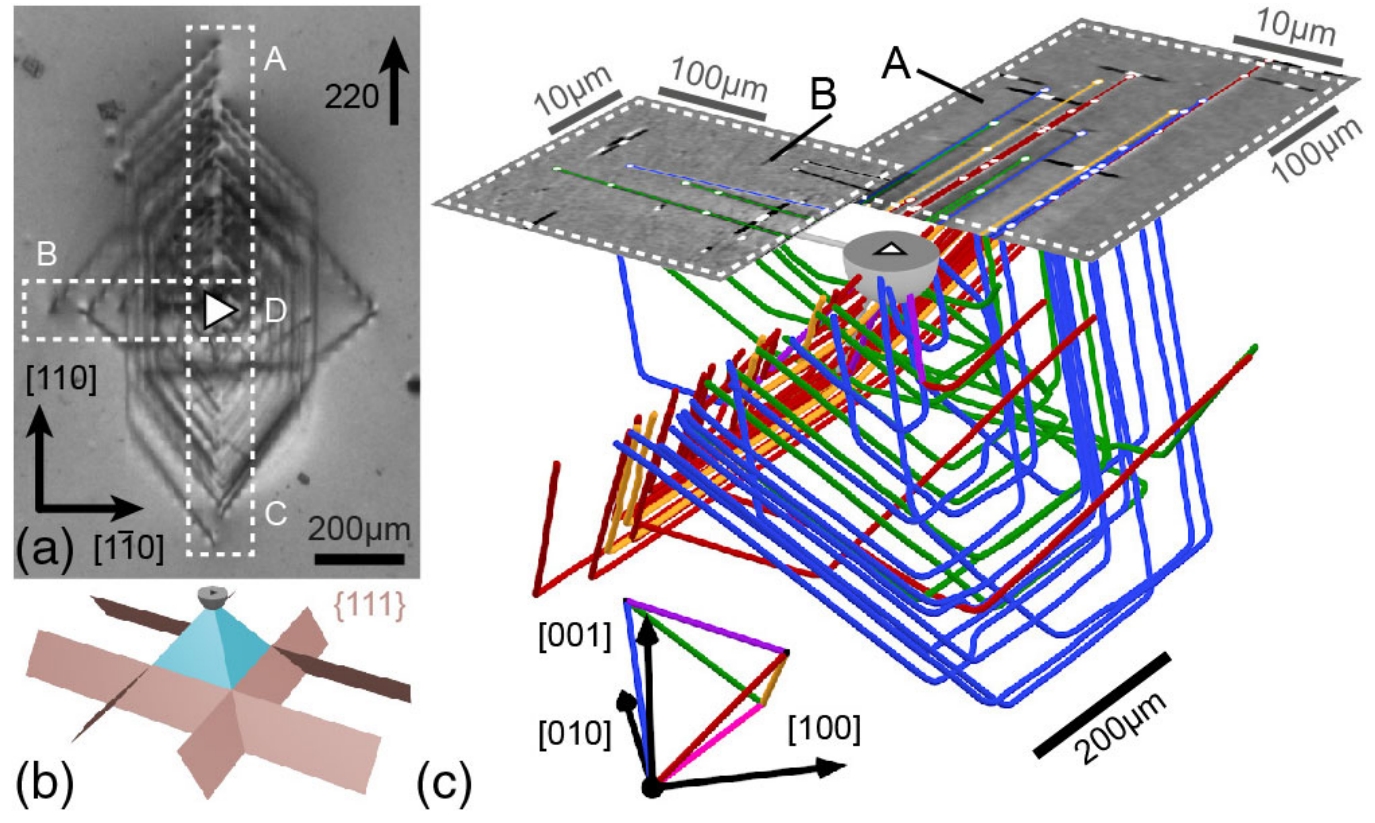
Combining X-ray diffraction laminography, X-ray white-beam topography and circularly polarized visible light differential interference contrast (CDIC) microscopy a complete 3D image of dislocation propagation as a result of mechanical damage (through indentation on the wafer surface, position indicated by the white triangle) can be constructed.
from: Correlated Three-Dimensional Imaging of Dislocations: Insights into the Onset of Thermal Slip in Semiconductor Wafers, Hänschke, D.; Danilewsky, A.; Helfen, L.; Hamann, E.; Baumbach, T.,2017. Physical review letters, 119 (21), Art. Nr.: 215504, doi:10.1103/PhysRevLett.119.215504 |
|
Methodical and instrumental development
Development and optimization of diffraction-based full-field X-ray imaging techniques, combining topography, full-field micro-diffraction imaging, diffraction laminography and tomography for correlative spatio-temporally resolved diffraction experiments. In combination with simulations (forward calculation of the measured diffraction contrast, FEM e.g. of thermal and mechanical stresses, dislocation dynamics, etc.) and complementary techniques such as µ-Raman spectroscopy and circular differential interference contrast microscopy (CDIC), the novel synchrotron methodology will enable a new level of comprehensive research studies of crystalline properties on technologically relevant length/time scales as well as under application-relevant conditions.
Link to external project page: https://www.strobos-code.org/
Publications
- Camattari R., Romagnoni M., Bandiera L., Bagli E., Mazzolari A., Sytov A., Haaga S., Kabukcuoglu M., Bode S., Hänschke D., Danilewsky A., Baumbach T., Bellucci V., Guidi V., and Cavoto G., X-ray Characterization of Self-standing Bent Si Crystal Plates for Large Hadron Collider Beam Extraction, Journal of Applied rystallography 53, 486-493 (2020)
- Asadchikov V., Buzmakov A., Chukhovsky F., Dyachkova I., Zolotov D., Danilewsky A., Baumbach T., Bode S., Haaga S., Hänschke D., Kabukcuoglu M., Balzer M., Caselle M., and Suvorov E., X-ray Topo-tomography Studies of Linear Dislocations in Silicon Single Crystals, Journal of Applied Crystallography 51(6), 1616-1622 (2018)
- Hänschke D., Hamann E., and Baumbach T., Neuartiger Röntgenblick auf Kristallversetzungen, Physik in unserer Zeit 49, 58-59 (2018)
- Faragó T., Mikulik P., Ershov A., Vogelgesang M., Hänschke D., and Baumbach T., syris: a flexible and efficient framework for X-ray imaging experiments simulation, Journal of Synchrotron Radiation 24, 1283-1295 (2017)
- Hänschke D., Danilewsky A., Helfen L., Hamann E., and Baumbach T., Correlated Three-Dimensional Imaging of Dislocations: Insights into the Onset of Thermal Slip in Semiconductor Wafers, Physical Review Letters 119, 215504 (2017) – Editors` Suggestion.
- Asadchikov V. E., Butashin A. V., Buzmakov A. V., Deryabin A. N., Kanevsky V. M., Prokhorov I. A., Roshchin B. S., Volkov Y. O., Zolotov D. A., Jafari A., Alexeev P., Cecilia A., Baumbach T., Bessas D., Danilewsky A. N., Sergueev I., Wille H.-C., and Hermann, R. P., Single-crystal sapphire microstructure for high-resolution synchrotron X-ray monochromators, Crystal Research and Technology 51, 290 (2016)
- Vogelgesang M., Farago T., Morgeneyer T. F., Helfen L., dos Santos Rolo T., Myagotin A., and Baumbach T., Real-time image-content-based beamline control for smart 4D X-ray imaging, Journal of Synchrotron Radiation 23, 1254-1263 (2016)
- Chilingaryan S., Shkarin A., Shkarin R., Vogelgesang M., and Tsapko S., Benchmark for FFT Libraries, Applied Mechanics and Materials 756, 673-677 (2015)
- Li Z. J., Danilewsky A. N., Helfen L., Mikulik P., Hänschke D., Wittge J., Allen D., McNally P., and Baumbach T., Local strain and defects in silicon wafers due to nanoindentation revealed by full-field X-ray microdiffraction imaging, Journal of Synchrotron Radiation, 22, 1083-1090 (2015)
- Rota L., Caselle M., Chilingaryan S., Kopmann A., and Weber M., A PCIe DMA Architecture for multi-Gigabyte per Second Data Transmission, IEEE Transactions on Nuclear Science 62, 972-976 (2015)
- Shkarin A., Ametova E., Kopmann A., Chilingaryan S., Shkarin R., Tsapko S., Dritschler T., and Vogelgesang M., An Open Source GPU Accelerated Framework for Flexible Algebraic Reconstruction at Synchrotron Light Sources, Fundamenta Informaticae 141, 259-274 (2015)
- Shkarin R., Ametova E., Chilingaryan S., Dritschler T., Kopmann A., Mirone A., Shkarin A., Tsapko S., and Vogelgesang M., GPU-optimized Direct Fourier Method for On-line Tomography, Fundamenta Informaticae 141, 245-258 (2015)
- Stevanovic U., Caselle M., Cecilia A., Chilingaryan S., Farago T., Gasilov S., Herth A., Kopmann A., Vogelgesang M., Balzer M., Baumbach T., and Weber M., A control system and streaming DAQ platform with image-based trigger for X-ray imaging, IEEE Transactions on Nuclear Science 62, 911-918 (2015)