
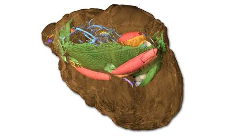
The rock (brown) preserved the beetle as a fossil. Ephemeral soft tissue and fragile limbs are represented excellently in three dimensions. (Photo: A. Schwermann / Th. van de Kamp / KIT)
Investigation of fossil arthropods like insects is largely restricted to external morphology, lacking information about internal characters which are crucial to establishing systematic position, ecological role and evolutionary trends. Moreover, the fossil record of arthropods containing internal characters is dominated by amber inclusions, which show representational bias.
By employing synchrotron X-ray microtomography at ANKA’s TOPO-TOMO beamline, a team led by scientists from the University of Bonn and KIT discovered well-preserved three-dimensional anatomy in 30-million-year old mineralized beetles from the fissure fillings in Quercy (south-central France), facilitating a detailed description of the species and its systematic relationship.
Despite being known for over a century, these fossils have been neglected for decades, mainly because of the poor condition of their outer surfaces. Modern X-ray imaging techniques now provided an incredibly detailed look at their internal structures. The results, which were published in the journal eLife, demonstrate that mineralized fossils, even those of apparently poor preservation, constitute a rich but yet largely unexploited source of anatomical data of fossil arthropods.
Reference:
Achim H. Schwermann, Tomy dos Santos Rolo, Michael S. Caterino, Günter Bechly, Heiko Schmied, Tilo Baumbach & Thomas van de Kamp: Preservation of three-dimensional anatomy in phosphatized fossil arthropods enriches evolutionary inference; “eLife”; DOI: 10.7554/eLife.12129