
HIGH-LIFE
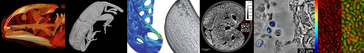
The main goal of the BMBF-funded HIGH-LIFE project is to enable the acquisition of complete 3D morphological information of small animals. The project addresses common requirements of important science cases in two fields of current biological research: (1) vertebrate model organisms such as fish, frog etc., providing indispensable paradigms to study development and disease, and (2) arthropods such as insects, which fulfill key functions in our ecosystems. Both cases share the need for morphological studies of whole organisms as well as its organs and tissues.
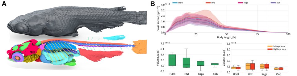
A: Anatomical atlas: segmented medaka with coloured organs. B. Correlation data: comparative morphometric analysis for four genetically distinct strains1.
A key objective of HIGH-LIFE is to establish dedicated experimental modalities for 3D digitization of small animal morphology by synchrotron radiation at PETRA III allowing advanced measurement, visualization, and, supported by artificial intelligence (AI), analysis of 3D morphological datasets. This includes instrumentation and methods for full-animal imaging, for hierarchical imaging down to cellular level and even for in vivo cine-tomography and time-lapse imaging. HIGH-LIFE builds on compiling various individual solutions and approaches, methods, instrumentation and software components developed in previous BMBF collaborative research projects (UFO I/II, CODE-VITA and NOVA) and combining them into smart and consistent morphological data acquisition, handling and analysis pipelines for small animals.
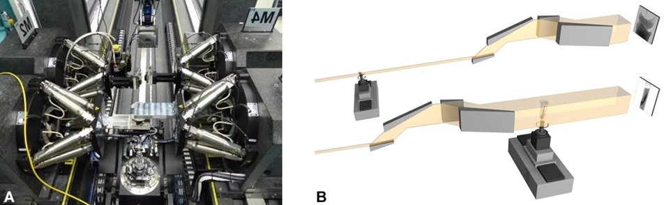
Bragg Magnifier instrumentation. A. BMM arrangement in a feasibility experiment at MIQA at KIT. B. Schematic configuration of BMM (top) and BMC (bottom) modes.
For HIGH-LIFE, the Bragg Magnifier crystal optics2,3 are a central feature, both in the context of dose-efficiency and the ability to vary the FoV. The BM systems can be used in two functionally different configurations:
- upstream of the sample, the Bragg Magnifier Beam Conditioner (BMC) conditions the size and coherence of the beam by either compression or expansion – in the latter case delivering a highly-parallel and monochromatic and therefore highly coherent output beam with strongly enlarged FoV.
- downstream of the sample, the Bragg Magnifier Microscope (BMM) acts as a very dose-efficient microscope with sub-µm resolution (resolution limit ~250 nm in case of Ge crystals).
The experimental techniques developed in the scope of HIGH-LIFE will pioneer hierarchical small animal imaging at PETRA III by implementing and integrating all necessary components, methods and analysis tools into the CODE-VITA experimental end station for the second experimental hutch at P23. Before transfer to PETRA III, all essential components will be commissioned at MIQA at KIT, and to a large extent tested at the first experimental hutch of P23. In consideration of a perspective storage ring PETRA IV, the components will be designed to be compatible to such an upgrade.
In summary, HIGH-LIFE will provide the user community with a dedicated experimental setup to enable generation of primary (raw), secondary (reconstructed) and tertiary (pre-analyzed) morphological data, which can later be correlated to complementary data such as genetic or environmental information. HIGH-LIFE aims to transfer these data into the Smart Repository for 3D Morphological Studies at Organisms of the Di-Morph Facility at the Science Data Center for Life Sciences (LifeSDC) presently planned at Heidelberg University. In this way, morphological data sets and correlated results will be made available to the international life science community and the general public.
Project Partners

| https://www.cos.uni-heidelberg.de/ | https://naturkundemuseum-bw.de/ |
Funding
HIGH-LIFE is funded by the BMBF as a joint-research project (Verbundprojekt) under the following numbers: 05K19VH1 (Univ. Heidelberg), 05K19VKE (KIT, LAS), 05K19VNA (Staatl. Museum für Naturkunde Stuttgart)
Publications
-
Evolution of flexible biting in hyperdiverse parasitoid wasps, Kamp, T. van de; Mikó, I.; Staniczek, A. H.; Eggs, B.; Bajerlein, D.; Faragó, T.; Hagelstein, L.; Hamann, E.; Spiecker, R.; Baumbach, T.; Janšta, P.; Krogmann, L., 2022. Proceedings of the Royal Society B: Biological Sciences, 289 (1967), Art.-Nr.: 2021.2086. doi:10.1098/rspb.2021.2086
-
Quantitative morphometric analysis of adult teleost fish by X-ray computed tomography
Weinhardt, V.; Shkarin, R.; Wernet, T.; Wittbrodt, J.; Baumbach, T.; Loosli, F., 2018. Scientific reports, 8 (1), Art. Nr.: 16531. doi:10.1038/s41598-018-34848-z -
In-line Bragg magnifier based on V-shaped germanium crystals, Vagovič, P., Korytár, D., Mikulík, P., Cecilia, A., Ferrari, C., Yang, Y., Hänschke, D., Hamann, E., Pelliccia, D., Lafford, T.A., Fiederle, M. & Baumbach, T., J. Synchrotron Rad. 18, 753-760 (2011).
-
T. High-resolution high-efficiency X-ray imaging system based on the in-line Bragg magnifier and the Medipix detector, Vagovič, P., Korytár, D., Cecilia, A., Hamann, E., Svéda, L., Pelliccia, D., Härtwig, J., Záprazný, Z., Oberta, P., Dolbnya, I., Shawney, K., Fleschig, U., Fiederle, M. & Baumbach, J. Synchrotron Rad. 20, 153-159 (2013).